
The Art and Science Behind Realistic 3D Printed Skulls and Anatomical Models
The intersection of medicine, education, and advanced manufacturing has never been more fascinating than in the world of 3D printed anatomical models. Of particular note are skulls and full anatomical models that replicate the intricacies of human anatomy with jaw-dropping realism. As a 3D printing expert, I’ve witnessed firsthand how these models are revolutionizing everything from surgical planning to classroom demonstrations.
The Role of 3D Printing in Anatomical Accuracy
Traditional anatomical models have long been used to teach students and aid professionals, but their limitations in detail, material, and customization have spurred demand for something better. Enter 3D printing—specifically, advanced techniques such as stereolithography (SLA), PolyJet, and multi-material fused deposition modeling (FDM), which have raised the bar for anatomical realism.
With access to DICOM files from CT or MRI scans, modern software can reconstruct patient-specific anatomy down to sub-millimeter accuracy. This digital data is then transformed into a printable file, allowing for the creation of skulls and bones that mirror the exact texture, density, and pathology of the real thing.
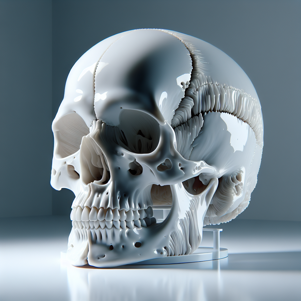
Material Choices for Hyper-Realism
The secret to the most realistic 3D printed skulls lies in material selection and post-processing. For bone-like translucency and density, resins and photopolymers such as those used in SLA and PolyJet are preferred. Some printers can even blend materials to simulate cortical and cancellous bone layers, producing models that not only look accurate but also feel correct during handling or surgical simulation.
For tactile feedback, composite filaments with ceramic or gypsum additives, or even calcium-phosphate-infused resins, can be used. These advanced materials allow for drilling and cutting, offering invaluable practice for surgeons and students alike.
Applications of Realistic Anatomical Models
- Medical Education: Life-like skulls help students understand complex structures—from cranial sutures to foramina—far more effectively than 2D images or generic models.
- Surgical Planning: Custom, patient-specific skulls empower surgeons to rehearse complex procedures, reducing risk and improving outcomes.
- Forensic Science: 3D printed replicas assist in facial reconstruction, trauma analysis, and court presentations, preserving the original specimen’s integrity.
- Museum and Display: Hyper-realistic models can be safely handled by the public, and even painted or aged for historical accuracy without risking irreplaceable artifacts.
Pushing the Limits: Full Color and Multi-Material Printing
With the advent of PolyJet and full-color binder jetting technologies, today’s anatomical models can accurately display not just the shape but also the coloration of real bone, soft tissue, and even pathology. This multi-color capability is a game-changer for teaching and diagnostics, allowing for clear visual differentiation between anatomical regions or disease states.
Post-Processing: The Final Touch for Authenticity
To achieve museum-quality realism, post-processing is critical. Techniques such as sanding, painting, staining, and clear coating are often employed. For skulls, artists may use subtle washes of ink or paint to highlight natural porosity and age, while dental structures can be polished for enamel shine.
Challenges and Future Directions
Despite remarkable advances, challenges remain—especially in simulating the full range of textures and mechanical properties found in real tissue. Hybrid models that combine rigid and flexible materials are already making headway, and ongoing research promises even greater anatomical fidelity.
As biocompatible and even bioresorbable materials enter the market, the gap between model and reality continues to shrink. In the near future, we may see 3D prints that not only mimic the look and feel of bone but can also host living cells for regenerative medicine.
Conclusion
The most realistic 3D printed skulls and anatomical models are more than just replicas—they are powerful tools that advance education, improve patient care, and unlock new possibilities for research and display. As technology continues to evolve, the boundary between model and reality will blur even further, ushering in a new era of anatomical understanding and innovation.
Leave a Reply